Conventional histology examines only a thin slice of a tumor, which is a fundamental problem for cancers like follicular thyroid carcinoma, where the critical diagnostic features (capsular and vascular invasion) can be focal and easily missed. This project develops micro-CT based tools to move beyond that limitation, offering a full 3D view of the tumor without any additional sample preparation beyond standard FFPE block embedding.

3D virtual histology
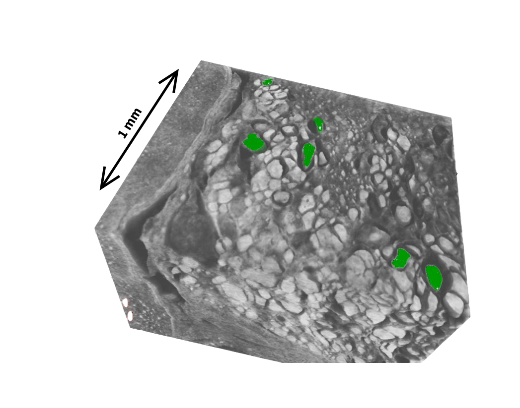
The first part of the project established micro-CT as a practical tool for 3D virtual histology of follicular thyroid carcinomas (FTC). We optimized the scanning protocol for FFPE tissue blocks and validated it on a cohort of 99 FTCs and 31 follicular adenomas. Capsular invasion was clearly detectable, and foci of possible vascular invasion could be flagged for targeted follow-up histology [1].
Key results:
- Positive predictive value: 100%
- Sensitivity: 86%, Accuracy: 89%
Imaging biomarker discovery
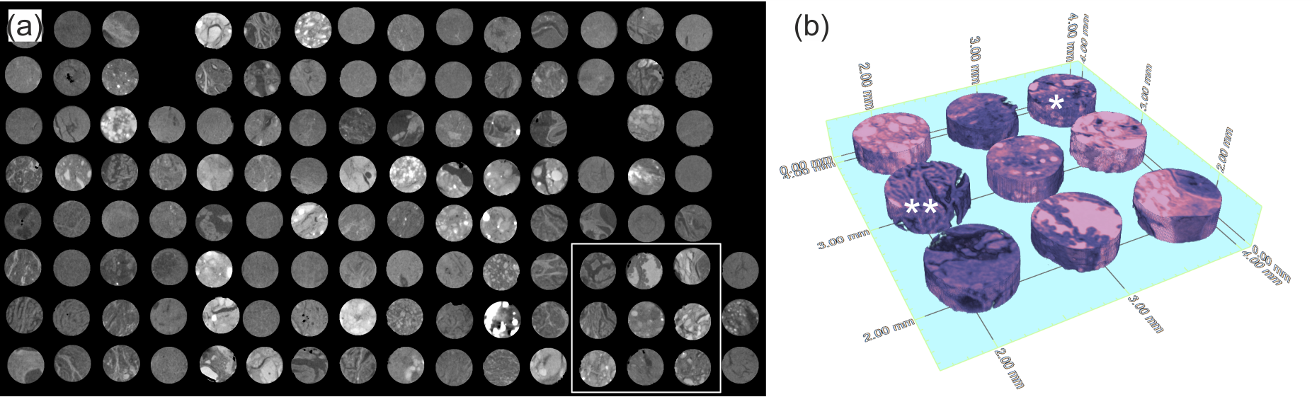
The second part asks a deeper question: can the 3D texture of a micro-CT scan encode molecular information about the tumor? Using radiomics on next-generation tissue microarrays (ngTMAs) from 418 patients, we extracted interpretable texture features and trained classifiers for several clinically relevant tasks. SHAP analysis was used to identify which textural patterns drove each classification decision, keeping the models transparent and interpretable.
Results at a glance:
- Neoplastic vs. non-neoplastic tissue: AUROC = 0.87 ± 0.03
- Papillary vs. follicular thyroid carcinoma: AUROC = 0.84 ± 0.05
- BRAF V600E mutation status: AUROC = 0.77 ± 0.06
- Preliminary signal for TERT promoter mutations: AUROC = 0.76 ± 0.15 [3]
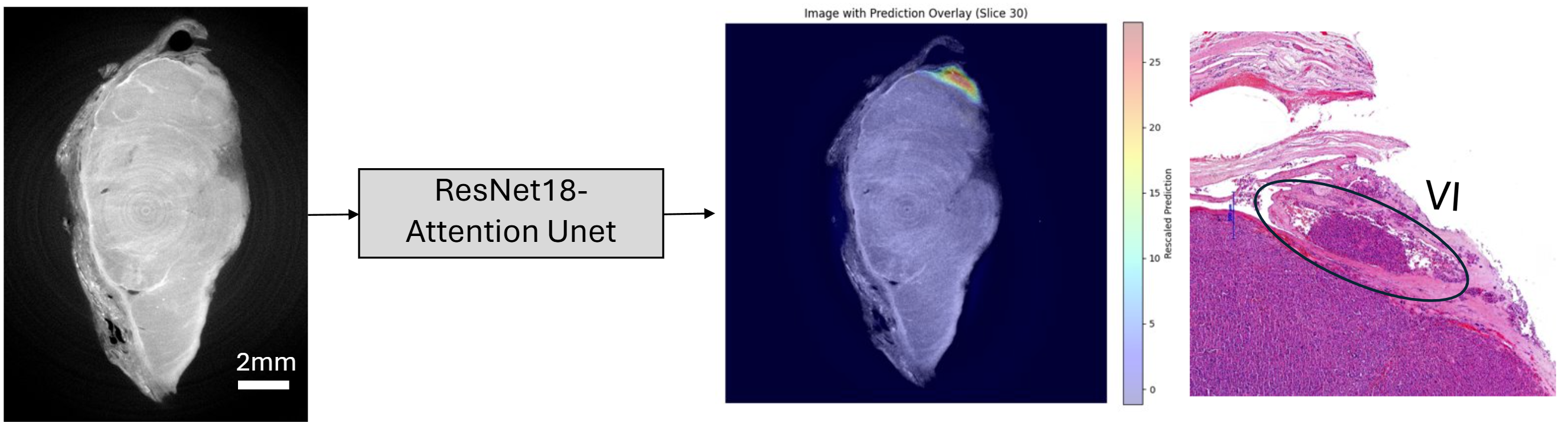
Automated attention map generation
An ongoing extension of the project is developing a deep learning model that automatically generates spatial attention maps highlighting regions of capsular and vascular invasion in FTCs, removing the need for manual inspection of the full 3D volume.
References
[1] Tajbakhsh, K., Stanowska, O., Neels, A., Perren, A., & Zboray, R. (2024). 3D virtual histopathology by phase-contrast X-ray micro-CT for follicular thyroid neoplasms. IEEE Transactions on Medical Imaging, 43(7), 2670-2678.
[2] Tajbakhsh, K., Stanowska, O., Bossart, J., Buljan, M., Neels, A., Mogl, M. T., … & Perren, A. (2025). Follicular thyroid carcinoma relapse cases-revisited by X-ray 3D virtual histology. Endocrine Pathology, 36(1), 46.
[3] Tajbakhsh, K., Stanowska, O., Buljan, M., Neels, A., Perren, A., & Zboray, R. (2025). Mapping 3D Heterogeneity of Thyroid Tumors Using Micro-CT based Radiomics. bioRxiv, 2025-06.